AUTOPSY REPORT
NAME: RAMSEY, JONBENET AUTOPSY NO:96A-155
DOB: 08/06/90 DEATH D/T: 12/26/96@ 1323
AGE: 6Y AUTOPSY D/T: 12/27/96 @0815
SEX: F ID NO: 137712
PATH MD: MEYER COR/MEDREC#: 1714-96-A
TYPE: COR
FINAL DIAGNOSIS:
I. Ligature strangulation
A. Circumferential ligature withassociated ligature furrow of neck
B. Abrasions and petechial hemorrhages,neck
C. Petechial hemorrhages, conjunctivalsurfaces of eyes and skin of face
II. Craniocerebral injuries
A. Scalp contusion
B. Linear, comminuted fracture ofright side of skull
C. Linear pattern of contusionsof right cerebral hemisphere
D. Subarachnoid and subdural hemorrhage
E. Small contusions, tips of temporallobes
III. Abrasion of right cheek
IV. Abrasion/contusion, posteriorright shoulder
V. Abrasions of left lower backand posterior left lower leg
VI. Abrasion and vascular congestionof vaginal mucosa
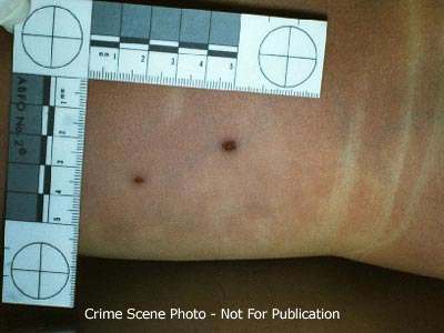
VII. Ligature of right wrist
TOXICOLOGIC STUDIES
blood ethanol - none detected
blood drug screen - no drugs detected
CLINICOPATHOLOGIC CORRELATION: Causeof death of this six year old female is asphyxia by strangulation associated with craniocerebral trauma.
John E. Meyer, M.D.
Pathologist
jn/12/27/96
NAME: RAMSEY, JONBENET AUTOPSY NO:96A-155 Page 2
The body of this six year old female was first seen by me after I was called to an address identified as 755 15th street in Boulder, Colorado, on 12/26/96. I arrived at the scene approximately 8 p.m. on 12/26 and entered the house where the decedent's body was located at approximately 8:20 p.m. I initially viewed the body in the living room of the house. The decedent was laying on her back on the floor, covered by a blanket and a Colorado Avalanche sweatshirt. On removing these two items from the top of the body the decedent was found to be lying on her back with her arms extended up over her head. The head was turned to the right. A brief examination of the body disclosed a ligature around the neck and a ligature around the right wrist. Also noted was a small area of abrasion or contusion below the right ear on the lateral aspect of the right cheek. A prominent dried abrasion was present on the lower left neck. After examining the body, I left the residence at approximately 8:30 p.m.
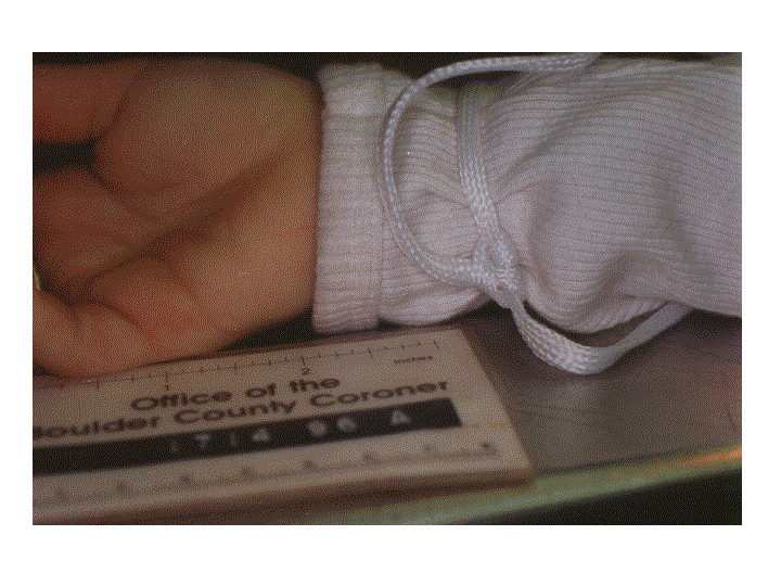
EXTERNAL EXAM: The decedent is clothed in a long sleeved white knit collarless shirt, the mid anterior chest area of which contains an embroidered silver star decorated with silver sequins.Tied loosely around the right wrist, overlying the sleeve of the shirt is a white cord. At the knot there is one tail end which measures 5.5 inches in length with a frayed end. The other tail of the knot measures 15.5 inches in length and ends in a double loop knot. This end of the cord is also frayed.
There are no defects noted in the shirt but the upper anterior right sleeve contains a dried brown-tan stain measuring 2.5 x 1.5 inches, consistent with mucous from the nose or mouth. There are long white underwear with an elastic waist band containing a red and blue stripe. The long underwear are urine stained anteriorly over the crotch area and anterior legs. No defects are identified. Beneath the long underwear are white panties with printed rose buds and the words "Wednesday" on the elastic waistband. The underwear is urine stained and in the inner aspect of the crotch are several red areas of staining measuring up to 0.5 inch in maximum dimension.
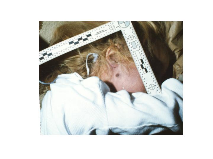
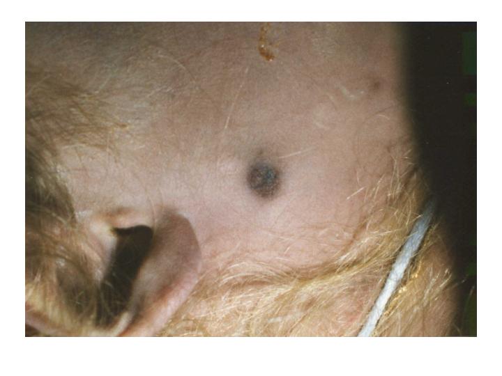
EXTERNAL EVIDENCE OF INJURY: Located just below the right ear at the right angle of the mandible, 1.5 inches below the right external auditory canal is a 3/8 X 1/4 inch area of rust colored abrasion.
In the lateral aspect of the left lower eyelid on the inner conjunctival surface is a 1 mm in maximum dimension petechial hemorrhage.Very fine, less than 1 mm petechial hemorrhages are present on the skin of the upper eyelids bilaterally as well as on the lateral left cheek. On everting the left upper eyelid there are much smaller, less than 1 mm petechial hemorrhages located on the conjunctival surface. Possible petechial hemorrhages located on the conjunctival surface. Possible petechial hemorrhages are also seen on the conjunctival surfaces of the right upper and lower eyelids, but livor mortis on this side of the face makes definite identification difficult.
Wrapped around the neck with a double knot in the midline of the posterior
Page 3
neck is a length of white cord similar to that described as being tied around the right wrist.
This ligature cord is cut on the right side of the neck and removed. A single black ink mark is placed on the left side of the cut and a double black ink mark on the right side of the cut. The posterior know is left intact. extending from the knot on the posterior aspect of the neck are two tails of the knot, one measuring 4 inches in length and having a frayed end, and the other measuring 17 inches in length with the end tied in multiple loops around a length of a round tan-brown wooden stick with measures 4.5 inches in length. This wooden stick is irregularly broken at both ends and there are several colors of paint and apparent glistening varnish on the surface. Printed in gold letters on one end of the wooden stick is the word "Korea." The tail end of another word extends from beneath the loops of the cord tied around the stick and is not able to be interpreted. Blonde hair is entwined in the knot on the posterior aspect of the neck as well as in the cord wrapped around the wooden stick. The white cord is flattened and measures approximately 1/4 inch in width. It appears to be made of a white synthetic material. Also secured around the neck is a gold chain with a single charm in the form of a cross.
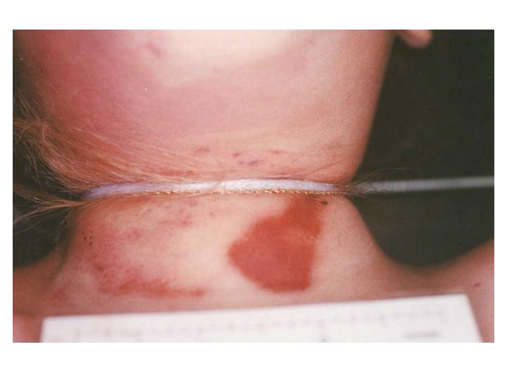
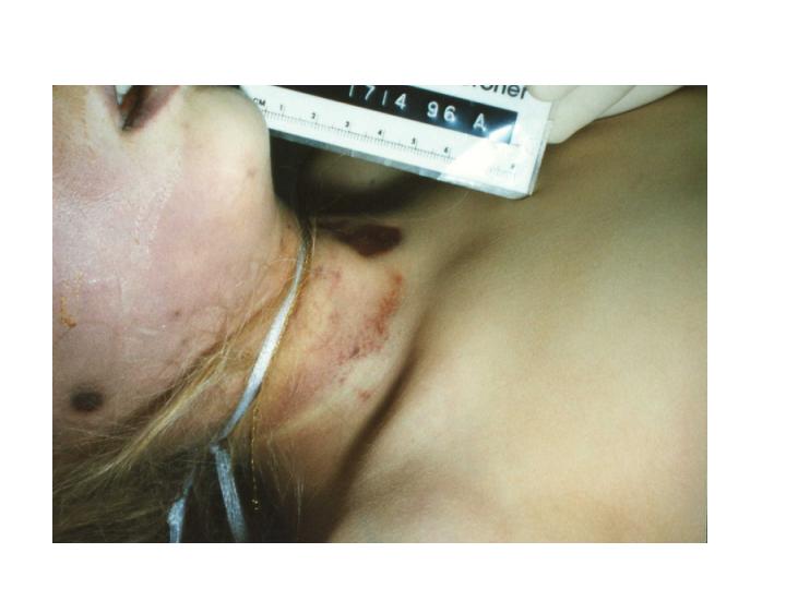
A deep ligature furrow encircles the entire neck. The width of the furrow varies from one-eighth of an inch to five/sixteenths of an inch and is horizontal in orientation, with little upward deviation. The skin of the anterior neck above and below the ligature furrow contains areas of petechial hemorrhage and abrasion encompassing an area measuring approximately 3 X 2 inches. The ligature furrow crosses the anterior midline of the neck just below the laryngeal prominence, approximately at the level of the cricoid cartilage. It is almost completely horizontal with slight upward deviation from the horizontal towards the back of theneck. The midline of the furrow mark on the anterior neck is 8 inches below the top of the head. The midline of the furrow mark on the posterior neckis 6.75 inches below the top of the head.
The area of abrasion and petechial hemorrhage of the skin of the anterior neck includes on the lower left neck, just to the left of the midline, a roughly triangular, parchment-liferust colored abrasion which measures 1.5 inches in length with a maximum width of 0.75 inches. This roughly triangular shaped abrasion is obliquely oriented with the apex superior and lateral. The remainder of the abrasions and petechial hemorrhages of the skin above and below the anterior projection of the ligature furrow are nonpatterned, purple to rust colored, and present in the midline, right, and left areas of the anterior neck. The skin just above the ligature furrow along the right side of the neck contains petechial hemorrhage composed of multiple confluent very small petechial hemorrhages as well as several larger petechial hemorrhages measuring up to one-sixteenth and one-eighth of an inch in maximum dimension. Similar smaller petechial hemorrhages are
Page 4
present on the skin below the ligature
furrow on the left lateral aspect of the neck. Located on the right side
of the chin is a three-sixteenths by one-eighth of an inch area of superficial
abrasion. On the posterior aspect of the right shoulder is a poorly demarcated,very
superficial focus of abrasion/contusion which is pale purple in color and
measures up to three-quarters by one-half inch in maximum dimension.
Several linear aggregates of petechial
hemorrhages are present in the anterior left should just above deltopectoral
groove. These measure up to one inch in length by one-sixteenth to one-eighth
of an inch in width. On the left lateral aspect of the lower back, approximately
sixteen and one-quarter inches and seventeen and one-half inches below
the level of the top of the head are two dried rust colored to slightly
purple abrasions.
The more superior of the two measures one-eighth by one-sixteenth of an inch and the more inferior measures three-sixteenths by one-eighth of an inch. There is no surrounding contusion identified. On the posterior aspect of the left lower leg, almost in the midline, approximately 4 inches above the level of the heel are two small scratch-life abrasions which are dried and rust colored. They measure one-sixteenth by less than on-sixteenth of an inch and one-eighth by less than one-sixteenth of an inch respectively.
On the anterior aspect of the perineum, along the edges of closure of the labia majora, is a small amount of dried blood. A similar small amount of dried and semifluid blood is present in the skin of the fourchette and the vestibule. Inside the vestibule of the vagina and along the distal vaginal wall is reddish hyperemia. This hyperemia is circumferential and perhaps more noticeable on the right side and posteriorly.The hyperemia also appears to extend just inside the vaginal orifice. A1 cm red-purple area of abrasion is located on the right posterolateral area of the 1 X 1 cm hymenal orifice. The hymen itself is represented bya rim of mucosal tissue extending clockwise between the 2 and 10:00 positions.The area of abrasion is present at approximately the 7:00 position and appears to involve the hymen and distal right lateral vaginal wall andpossibly the area anterior to the hymen. On the right labia majora is a very faint area of violet discoloration measuring approximately one inch by three-eighths of an inch. Incision into the underlying subcutaneous tissue discloses no hemorrhage. A minimal amount of semiliquid thin watery red fluid is present in the vaginal vault. No recent or remote anal or other perineal trauma is identified.
REMAINDER OF EXTERNAL EXAMINATION:The unembalmed, well developed nourished caucasian female body measures 47 inches in length and weighs an estimated 45 pounds. The scalp is covered by long blonde hair which is fixed in two ponytails, one on top of the head secured by a cloth hair tie and blue elastic band, and one in the lower back of the head secured by a blue elastic band. No scalp trauma is identified. The external auditory canals are patent and free of blood.The eyes are green and the pupils
Page 5
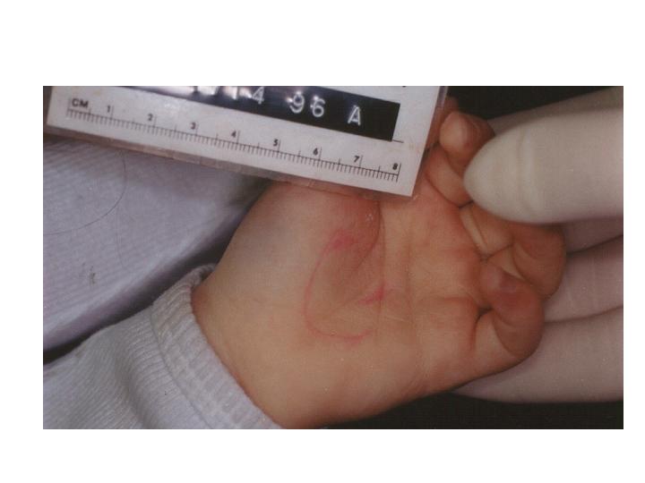
equally dilated. The sclerae are white. The nostrils are both patent and contain a small amount of tan mucous material. The teeth are native and in good repair. The tongue is smooth, pink-tan and granular. No buccal mucosal trauma is seen. The frenulum is intact. There is a slight drying artifact of the tip of the tongue. On the right cheek is a pattern of dried saliva and mucous material which does not appear to be hemorrhagic. The neck contains no palpable adenopathy or masses and the trachea and larynx are midline. The chest is symmetrical. Breasts are prepubescent. The abdomen is flat and contains no scars. No palpable organ omegaly or masses are identified. The external genitalia are that of a prepubescent female. No pubic hair is present. The anus is patent. Examination of the extremities is unremarkable. On the middle finger of the right hand is a yellow metal band. Around the right wrist is a yellow metal identification bracelet with the name "JonBenet" on one side and the date "12/25/96" on the other side. A red ink line drawing in the form of a heart is located on the palm of the left hand.
The fingernails of both hands are of sufficient length for clipping. Examination of the back is unremarkable. There is dorsal 3+ to 4+ livor mortis which is nonblanching. Livor mortis is also present on the right side of theface. At the time of the initiation of the autopsy there is mild 1 to 2+rigor mortis of the elbows and shoulders with more advanced 2 to 3+ rigor mortis of the joints of the lower extremities.
INTERNAL EXAM: The anterior chest musculature is well developed. No sternal or rib fractures are identified.
Mediastinum: The mediastinal contents are normally distributed. The 21 gm thymus gland has a normal external appearance. The cut sections are finely lobular and pink-tan. No petechial hemorrhages are seen. The aorta and remainder of the mediastinal structures are unremarkable.
Body Cavities: The right and leftthoracic cavities contain approximately 5 cc of straw colored fluid. The pleural surfaces are smooth and glistening. The percardial sac contains 3-4 cc of straw colored fluid and the epicardium and pericardium are unremarkable.The abdominal contents are normally distributed and covered by a smooth glistening serosa. No intraabdominal accumulation of fluid or blood is seen.
Lungs: The 200 gm right lung and175 gm left lung have a normal lobar configuration. An occasional scattered subpleural petechial hemorrhage is seen on the surface of each lung. The cut sections of the lungs disclose an intact alveolar architecture witha small amount of watery fluid exuding from the cut surfaces with mildpressure. The intrapulmonary bronchi and vasculature are unremarkable. No evidence of consolidation is seen.
Heart: The 100 gm heart has a normal external configuration. There are scattered subepicardial petechial hemorrhages over the anterior surface of
Page 6
the heart. The coronary arteries are normal in their distribution and contain no evidence of atherosclerosis.The tan-pink myocardium is homogeneous and contains no areas of fibrosisor infarction. The endocardium is unremarkable. The valve cusps are thin,delicate and pliable and contain no vegetation or thrombosis. The major vessels enter and leave the heart in the normal fashion. The foramen ovaleis closed.
Aorta and Vena Cava: The aorta is patent throughout its course as are its major branches. No atherosclerosisis seen. The vena cava is unremarkable.
Spleen: The 61 gm spleen has a finely wrinkled purple capsule. Cut sections are homogeneous and disclose readily identifiable red and white pulp. No intrinsic abnormalities are identified.
Adrenals: The adrenal glands are of normal size and shape. A golden yellow cortex surmounts a thin brown-tan medullary area. No intrinsic abnormalities are identified.
Kidneys: The 40 gm right kidney and 40 gm left kidney have a normal external appearance. The surfaces are smooth and glistening. Cut sections disclose an intact corticomedullary architecture. The renal papillae are sharply demarcated. The pelvocaliceal system is lined by gray-white mucosa which is unremarkable. Both uretersare patent throughout their course to the bladder.
Liver: The 625 gm liver has a normal external appearance. The capsule is smooth and glistening. Cut sections disclose an intact lobular architecture with no intrinsic abnormalities identified.
Pancreas: The pancreas is of normal size and shape. Cut sections are finely lobular and tan. No intrinsic abnormalitiesare identified.
Bladder: The bladder is contracted and contains no urine. The bladder mucosa is smooth and tan-gray. No intrinsic abnormalities are seen.
Genitalia: The upper portions ofthe vaginal vault contain no abnormalities. The prepubescent uterus measures3 X 1 X 0.8 cm and is unremarkable. The cervical os contains no abnormalities. Both fallopian tubes and ovaries are prepubescent and unremarkable by gross examination.
Gallbladder: The gallbladder contains 2-3 cc of amber bile. No stones are identified and the mucosa is smooth and velvety. The cystic duct, right and left hepatic duct and common bile duct are patent throughout their course to the duodenum.
G.I. Tract: The esophagus is empty.It is lined by gray-white mucosa. The
Page 7
stomach contains a small amount (8-10 cc) of viscous to green to tan colored thick mucous material without particulate matter identified. The gastric mucosa is autolyzed but contains no areas of hemorrhage or ulceration. The proximal portion of the small intestine contains fragmented pieces of yellow to light green-tan apparent vegetable or fruit material which may represent fragments of pineapple. No hemorrhage is identified. The remainder of the small intestine is unremarkable. The large intestine contains soft green fecal material. The appendix ispresent.
Lymphatic System: Unremarkable
Musculoskeletal System: Unremarkable
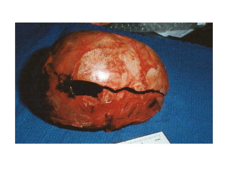
Skull and Brain: Upon reflection of the scalp there is found to be an extensive area of scalp hemorrhage along the right temporoparietal area extending from the orbital ridge, posteriorly all the way to the occipital area. This encompasses an area measuring approximately 7 X 4 inches. This grossly appears to be a fresh hemorrhage with no evidence of organization. At the superior extension of this area of hemorrhage is a linear to comminuted skull fracture which extends from the right occipital to posteroparietal area forward to the right frontal area across the parietal portion of the skull. In the posteroparietal area of this fracture is a roughly rectangular shaped displaced fragment of skull measuring one and three-quarters by one-half inch. The hemorrhage and the fracture extend posteriorly just past the midline of the occipital area of the skull. This fracture measures approximately 8.5 inches in length.
On removal of the skull cap there is found to be a thin film of subdural hemorrhage measuring approximately 7-8 cc over the surface of the right cerebral hemisphere and extending to the base of the cerebral hemisphere.The 1450 gm brain has a normal overall architecture. Mild narrowing of the sulci and flattening of the gyri are seen. No inflammation is identified.There is a thin film of subarachnoid hemorrhage overlying the entire right cerebral hemisphere. On the right cerebral hemisphere underlying the previously mentioned linear skull fracture is an extensive linear area of purple contusion extending from the right frontal area, posteriorly along the lateral aspect of the parietal region and into the occipital area. This area of contusion measures 8 inches in length with a width of up to 1.75 inches. At the tip of the right temporal lobe is a one-quarter by one-quarter inch similar appearing purple contusion. Only very minimal contusion is present at the tip of the left temporal lobe. This area of contusion measures only one-half inch in maximum dimension. The cerebral vasculature contains no evidence of atherosclerosis. Multiple coronal sections of the cerebral hemispheres, brain stem and cerebellum disclose no additional abnormalities. The areas of previously described contusion are characterized by purple linear streak-like discolorations of the gray matter perpendicular to the surface of the cerebral cortex. These extend approximately 5 mm into the
Page 8
cerebral cortex. Examination of the base of the brain discloses no additional fractures.
Neck: Dissection of the neck is performed after removal of the thoracoabdominal organs and the brain. The anterior strap musculature of the neck is serially dissected. Multiplesections of the sternocleidomastoid muscle disclose no hemorrhages. Sectionsof the remainder of the strap musculature of the neck disclose no evidence of hemorrhage. Examination of the thyroid cartilage, cricoid cartilage and hyoid bone disclose no evidence of fracture or hemorrhage. Multiple cross sections of the tongue disclose no hemorrhage or traumatic injury.The thyroid gland weighs 2 gm and is normal in appearance. Cut sections are finely lobular and red-tan. The trachea and larynx are lined by smooth pink-tan mucosa without intrinsic abnormalities.
MICROSCOPIC DESCRIPTION: (All sectionsStained with H&E)
(Slide key(A) scalp hemorrhage ,(B) sections of vaginal mucosa with smallest fragment representing area of abrasion at 7:00 position , (C) heart , (D-F) lungs , (G) liver andspleen , (H) pancreas and kidney , (I) thyroid and bladder , (J) thymusand adrenals , (K-L) reproductive organs , (M) larynx , (N-T) brain.
Myocardium: Sections of the ventricularmyocardium are composed of interlacing bundles of cardiac muscle fibers. No fibrosis or inflammation are identified.
Lungs: The alveolar architectureof the lungs is well preserved. Pulmonary vascular congestion is identified.No intrinsic abnormalities are seen.
Spleen: There is mild autolysisof the spleen. Both red-and-white pulp are identifiable.
Thyroid: The thyroid gland is composed of normal-appearing follicles. An occasional isolated area of chronic interstitial inflammatory infiltrate is seen. There is also a small fragment of parathyroid tissue.
Thymus: The thymus gland retains the usual architecture. The lymphoid material is intact and scattered Hassall'scorpuscles are identified. Mild vascular congestion is identified.
Trachea: There is mild chronic inflammationin the submucosa of the trachea.
Liver: The lobular architectureof the liver is well preserved. No inflammation or intrinsic abnormality are identified.
Page 9
Pancreas: There is autolysis ofthe pancreas which is otherwise unremarkable. Kidney: The overall architectureof the kidney is well preserved. There is perhaps mild vascular congestionin the cortex but no inflammation is identified.
Bladder: The transitional epithelium of the bladder is autolyzed. No significant intrinsic abnormalities areseen.
Reproductive Organs: Sections ofthe uterus are consistent with the prepubescent age. The ovary is unremarkable.
Adrenal: The architecture of the adrenal is well preserved and no intrinsic abnormalities are seen.
Brain: Sections from the areas ofcontusion disclose disrupted blood vessels of the cortex with surrounding hemorrhage. There is no evidence of inflammatory infiltrate or organization of the hemorrhage. Subarachnoid hemorrhage is also identified. Cortical neurons are surrounded by clear halos, as are glial cells.
Vaginal Mucosa: All of the sections contain vascular congestion and focal interstitial chronic inflammation.The smallest piece of tissue, from the 7:00 position of the vaginal wall/hymen, contains epithelial erosion with underlying capillary congestion. A small number of red blood cells is present on the eroded surface, as is birefringent foreign material. Acute inflammatory infiltrate is not seen.
EVIDENCE: Items turned over to theBoulder Police Department as evidence include: Fibers and hair from clothing and body surfaces; ligatures; clothing; vaginal swabs and smears; rectal swabs and smears; oral swabs and smears; paper bags from hands; fingernail clippings; jewelry; paper bags from feet; white body bag; samples of head hair, eyelashes and eyebrows; swabs from right and left thighs and right cheek; red top and purple top tubes of blood.
END OF REPORT
Note from Dr. John Meyer August13, 1997
Contrary to several media reports over the past few days, the autopsy report on JonBenet Ramsey does not and has never contained information on the estimated time of death. I have not been able to determine the original source of the statement that the report contained the estimated time of death, but it certainly did not come from this office.
The time of an "unwitnessed" death is very difficult to determine with any precision, and at best is an estimate based not only on autopsy findings but also on investigative information.
I consider estimation of time of death to be an interpretive finding rather than a factual statement, and it is not this Office's practice to include this estimate as part of any autopsy report. As has been stated in the past, it would also be inappropriate for me, as a potential expert and material witness, to make interpretive statements prior to testifying in court.
John E. Meyer, M.D.
Boulder County Coroner
